X-Light V2tp
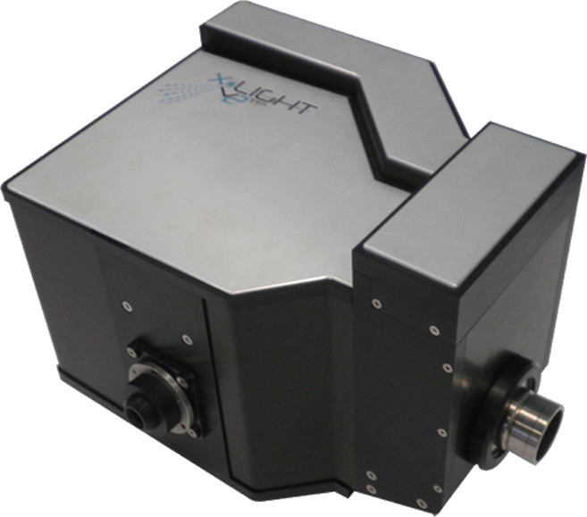
Confocal imaging system from Crest Optics
The X-Light V2tp is a unique system for spinning disk confocal imaging. It features unparalleled versatility:
- Compatibility with all major inverted and upright microscopes
- Plug‐in spinning disk box, allowing a change of pinhole pattern in seconds
- Use of custom relay lenses for mitigation of aberrations of of a large field of view
- Utilization of full field of view in sCMOS cameras
- Full compatibility with Crest Optics Video Confocal Super‐resolution module (“VCS”) for 3-D resolution enhancement
- Movable disk for widefield imaging

The X-Light V2tp Confocal Imager
The X-Light V2tp Confocal Imager is a full-spectrum spinning disk confocal imager that attaches to most major models of inverted or upright fluorescence microscope. It is designed to utilize the latest imaging technologies and is ideal for live cell imaging in biological applications.
The X-Light V2tp Confocal Imager allows imaging at the extremes of performance of a current‐generation scientific camera, and it provides for objectives used when imaging with a camera with a smaller detector. It takes advantage of laser launches and LED light sources.
The result is a flexible confocal add‐on for existing microscopes that brings the latest commercialized technologies to microscopy. The X-Light V2tp Confocal Imager is an extraordinary combination of capability and price.
Features
Acquisition Modes
A rapid motorized Disk In/DiskOut control provides access to widefield and confocal imaging modes. Switching between modes is automated by imaging software.
Nipkow Spinning Disk
A proprietary disk pattern design provides high confocal resolution, improved out‐of‐focus rejection and higher ratio of signal‐to‐noise. The result is < 800 nm confocal resolution with 60x 1.4 N.A. oil immersion objective.
Disks are available with pinholes sized for best results from different objectives:
- 40–μm holes, for objectives with N.A. < 1;
- 70–μm holes, for objectives with N.A. > 1;
- Custom pinhole diameters are available.
Disks are available to best suit particular camera setups.
- Single Hole Pattern: 22×22–μm field of view for large chips of scientific CMOS cameras;
- Dual Hole Patterns: 12×12–μm field of view for each pattern, for flexibility for smaller chips of CCD cameras.
Motor
The disk rotates at 15,000 revolutions per minute, providing excitation and imaging at speeds of current cameras.
Illumination Subsystems
- The excitation mount accomodates LED light sources or multimode fiber for lasers; its SMA connector provides high‐efficiency coupling;
- The gimbal mount for excitation eases alignment to produce the highest ratio of signal to noise;
- The motorized five‐position dichroic filter wheel and eight‐position emission filter wheel provide for automation and ease of interactive control.
Other optics
Custom relay lenses mitigate aberrations of of a large field of view.
Ease of installation and maintenance
The X-LIGHT V2tp addresses difficulties of both initial set-up and continuing maintenance of a confocal imager.
- The gimbal mount for excitation eases alignment that is required for maximal ratio of signal to noise;
- Adjustment of focus is accomplished without moving the camera;
- Fitler extraction tools ease insertion and removal of dichroic filters and emission filters.
VCS: Video Confocal Super-resolution
The VCS (“Video Confocal Super‐resolution”) module is an add‐on for the X-Light V2.
Patented Approach with High‐Speed Processing
CrestOptics VCS enhances the 3‐D resolution of the standard light microscope. It performs a two‐dimensional scan of the sample with a pinhole pattern and applies multiple algorithms to process the the emission, producing the image. The VCS technology is patented.
To speed the extensive calculation required, the VCS module uses CUDA technology from NVidia. The result is very fast processing.
Multiple Modes of Operation
The addition of the VCS module to the X-Light V2 provides a confocal imager with three modes of operation: Widefield Mode, Confocal Mode, and Super-resolution Mode.
Principles of Operation with VCS
VCS (“Video Confocal Super‐resolution”) is based on narrow-field illumination using scanning patterns and wide-field collection of raw images.
Detection algorithms already developed are able to super-resolve 3‐D structures in both compact and sparse specimens.
Although other techniques proposed and industrially developed mainly dedicate their efforts in extracting information from a relatively low spatial frequency range, VCM detection methods harness non-linear calculations exploiting the tops more than the belly of the raw signal intensity, collected as a function of illumination and detection positional parameters.
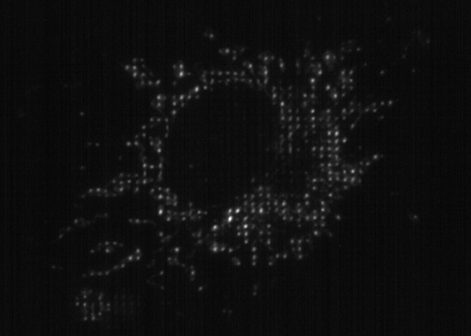
The figure below shows the schematic diagram for a basic VCS microscopy system. In this set‐up, the light source is focused onto a specified pattern that is optically conjugated to the microscope conjugate focal plane through a relay system. The mask is moved in the optical path with an X‐Y piezo motor system at each camera acquisition. The emission is collected widefield, and extensive processing performed to produce the final image.

The figure below shows a typical illumination pattern projected onto a sample.

L-FOV: Large Field of View
X-Light V2 L-FOV is the X-Light V2 variant which covers the latest very large field of view cameras introduced in the biological market.
Specifically customized on the Nikon Instruments Eclipse Ti2 microscope, it guarantees illumination uniformity, high-speed confocal acquisition and high optical imaging quality in a 25mm field of view.
X-Light V2 L-FOV delivers:
- Up to 25mm Field of View
- Illumination uniformity: drop-off corner to center <15%
- Confocal resolution with 60x NA 1.4 oil immersion Objective: <650nm
- Adapter mount for Eclipse Ti2 side port
- F-mount for camera mount
Technical Specifications for X-Light V2tp
| Feature | Detail |
| Disk Configurations Available | Dual Pinhole Patterns, for flexibility in matching smaller detectors with a single disk |
| Single Pinhole Pattern, for maximum field of view | |
| Acquisition Modes | Widefield, with fast motorized disk‐in, disk‐out |
| Confocal, for disk with single pinhole pattern | |
| Confocal, for each pattern of dual‐pattern disk | |
| Detector Field of View | Dual‐pattern disk: 10×10 mm (14 mm diagonal) |
| Single‐pattern disk: up to 22 mm diagonal | |
| Pattern Configuration | CrestOptics proprietary disk pattern design for high confocal resolution, improved rejection of Out‐of‐Focus information and higher S/N |
| Confocal Resolution with 60×/1.40 NA oil immersion objective: <650nm | |
| 40‐micron pinholes | |
| 50‐micron pinholes | |
| 60‐micron pinholes | |
| 70‐micron pinholes | |
| Custom spiral patterns available upon request | |
| Custom relay lenses | Diffraction‐limited across entire field of view when using high‐magnification high-N.A. objectives or low‐magnification low-N.A. objectives |
| Spinning Disk Rotation Speed | 15,000 RPM |
| Excitation mount | Gimbal mount, for easy alignment with optical axis of microscope and for best S/N |
| Dichroic Filter Wheel | 5 positions, motorized |
| Emission Filter Wheel | 8 positions, motorized |
| Ease of Installation | Plug‐in Spinning Disk |
| Includes tools for insertion and extraction of dichroic filters and emission filters | |
| Provides for focusing of detector focal plane without moving the detector | |
| Includes adapter for your choice of excitation: multimode laser or LED SMA 905 fiber |
Specifications for VCS (“Video Confocal Super‐resolution”)
- Standard and custom hole pattern
- Patterns differ from one other in hole diameter and spacing, best choice depends on microscope configuration and on sample characteristics
- Two patterns on one physical mask
- Pre‐aligned module with integrated LED or LASER launcher
- Bypass mode for widefield illumination and acquisition
- Up to 18 mm field of view
- LED and LASER source compatible
- Piezo motors speed structured illumination scan
- Motorized control for VCS pattern focusing and color correction
- Motorized mask positioning from VCS mode to Widefield/Confocal mode
- Lateral (X–Y) resolution ca. 120 nm measured
- limited by properties of optics, by objective magnification, and by camera pixel size
- Axial (Z) resolution ca. 300 nm
- Compatible objectives: highly-corrected immersion 60x and 100x
- 3‐D axial range: tested up to 110 micron
- Time resolution: ~5 sec/frame calculation included
- Camera requirements
- 6.5 µm pixel recommended for sCMOS camera
- sCMOS camera; QE>80% and low readout noise ≤ 1e- strongly recommended
Video card/CUDA Requirements
- Recommended for Full Performance:
- Compute Capability: Version 5.0 or higher
- GPU Memory: 4 GB or more
VCS/SCSR Pixel Calibration Table
| Camera Pixel Size | |||
| Magnification | 6.5 µm | 11 µm | 13 µm |
| 60× | 54.2 nm per pixel | 91.6 nm per pixel | 108 nm per pixel |
| 100× | 32.5 nm per pixel | 55 nm per pixel | 65 nm per pixel |
Images

Stack projection of HeLa cells with Qdot™ Conjugates
Nucleus – Qdot™ 655; Golgi – Qdot™ 585; Microtubules – Qdot™ 525.
Quantum Dot Corporation, Hayward, CA.

Stack projection of mouse intestine section
Labeled with Alexa Fluor® 350 WGA, Alexa Fluor® 568 phalloidin, and SYTOX® Green.
Molecular Probes/Invitrogen, Eugene, OR.

Maximum projection created from confocal sections through a sea urchin embryo
Embryo fixed and stained with anti-tubulin (green) and phalloidin (red)
Tubulin (green), Actin (red), (60× 1.4 N.A.)
Dr. George von Dassow, Center for Cell Dynamics, Friday Harbor Labs, University of Washington, Friday Harbor, WA.

Orthogonal (X–Z) view created from confocal sections through a sea urchin embryo
Embryo fixed and stained with anti-tubulin (green) and phalloidin (red)
Tubulin (green), Actin (red), (60× 1.4 N.A.)
Dr. George von Dassow, Center for Cell Dynamics, Friday Harbor Labs, University of Washington, Friday Harbor, WA.

3‐D reconstruction of imaged skin sample
Labeled using pan‐neuronal marker, protein gene product 9.5 localized with Cy3 and basement membrane marker, type IV collagen with Cy2. Endothelial cells are stained using Cy5‐Ulex europeaus Agglutinin Type 1.
Dr. William R. Kennedy and Gwen Wendelschafer-Crabb, University of Minnesota, Minneapolis, MN

Human Skin, maximum projection.
Basement membrane stained using Cy2, Neuron with Cy3, Endothelial cells with Cy5.
Dr. William R. Kennedy and Gwen Wendelschafer-Crabb, University of Minnesota.

Drosophila Embryo, Tubulin stained with GFP.
Tubulin stained with GFP.
Dr. Garrett Odell, Friday Harbor, University of Washington.

Time‐lapse images at a single confocal plane of a sand dollar embryo during mid‐blastula stage
Rhodamine‐tubulin and Alexa 488 phalloidin, Molecular Probes (Eugene, Oregon)
Dr. George von Dassow, Bill Bement, Center for Cell Dynamics, Friday Harbor Labs, University of Washington, Friday Harbor, WA

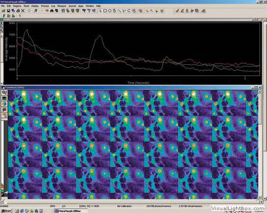
Time‐lapse recordings at 50 frames per second of calcium sparks in muscle cells
Captured using the Roper Cascade 512B CCD camera in conjunction with Metamorph® imaging software (Molecular Devices, Sunnyvale, CA)

Mouse Brain Confocal Performance Print
60 μm‐thick section of an entire mouse brain. The sample is stained with 4 colors (3 antibodies and DAPI) and has been scanned by TissueFAXS Confocal at 20x magnification factor. 1400 images per each confocal plane and a total of 29400 images for 21 layers z-stack.
Courtesy of TissueGnostics. Acquisitions by courtesy of Janelia Farm Research Campus, Howard Hughes Medical Institute.


