VisiView
High‐Performance Imaging Software from Visitron Systems GmbH
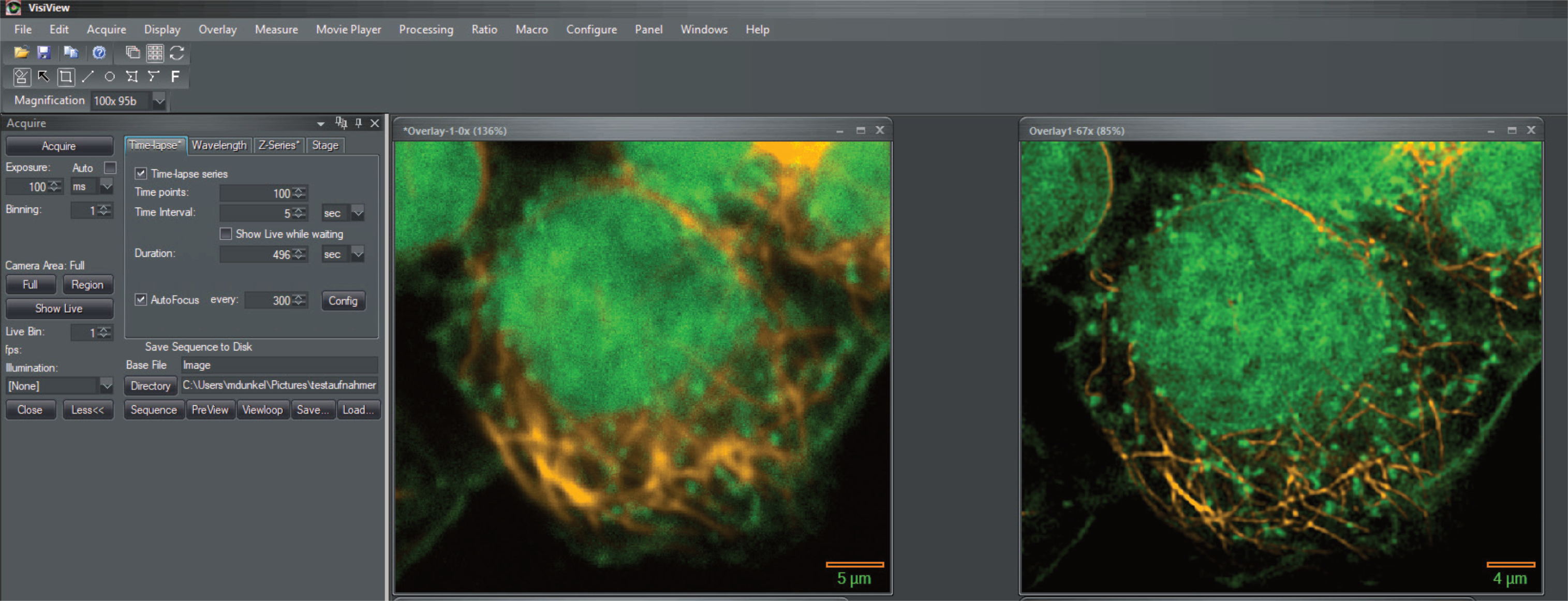
VisiView® is a high performance imaging software from Visitron Systems GmbH for bioimaging applications. It is designed to meet the needs for high‐speed image acquisition and processing with ease of use. It controls complex automated microscopes and microscope equipment in combination with multidimensional acquisition and analysis. Its multitasking ability supports simultaneous image acquisition and analysis. VisiView software represents the philosophy of simple operations and seamless integration of applied standards.
Life Science Imaging for the 2020s
VisiView is image acquisition, processing, measurement, and analysis software. it directly supports the acquisition of images and sequences of images as needed for today's research, from basic documentation to the most complex protocols requring the most exacting control of cameras, light sources, microscope automation, and add-on hardware. Sophisticated imaging processes or simple routines can be automated down to the push of a single button. Fully integrated options easily automate microscope control. VisiView includes tools needed to build imaging, analysis, and automated solutions for the laboratory. Extensions (such as Real-time 3-D Deconvolution and VirTex Realtime Experiment Control) smoothly integrate new processing, analysis, and hardware control into VisiView. VisView is a proven solution, built to take advantage of the technologies brought to life science imaging in the 21st Century.
It addresses these complex protocols:
- Time‐lapse image acquisition;
- Single or multi‐channel acquisition;
- Multi‐dimensional time‐lapse acquisition with display of overlayed images;
- High‐speed image acquisition (“stream acquisition”), including synchronization of high-speed focus and laser emission with minimized photobleaching;
- Control of motorized X‐Y stage;
- Scanning of multi‐well plates or entire slides.
Formats for saved images that give you full access to your data
VisiView saves images in the user’s choice of OME-TIFF files, TIFF files, or STK files. The OME-TIFF format is an open standard developed by the Open Microscopy Environment consortium and supported by Fiji, ImageJ as well as commercial applications.
Image Display
VisiView provides access to a full suite of image display features and tools. With VisiView, you can easily combine multiple image sequences and readily blend as many as seven illumination settings, combining grayscale ( e.g. DIC) images with fluorescence image sequences. VisiView's Five‐Dimensional Visualization option gives you the ability to visualize through-focus image stacks, adding to that the replay of time sequences and display of the color overlaay; it also provides for making measurements.
Image Processing
The processing dialog of VisiView provides background subtraction and shading correction, noise reduction, and low pass filters. Additional processing features are available through optional VisiView modules:
- Real‐time 2–D Deconvolution;
- Real‐time 3–D Deconvolution;
- Feature enhancement via Artificial Intelligence filters;
- Image stitching — the creation of a large-format image from a set of images scanned using an X-Y stage.
Further features are accesible by means of IronPython macros; macros are provided that call OpenCVSharp to gain access to OpenCV routines.
Automation
VisiView provides repeatability through automation:
- Via acquisition setup files. An acquisition protocol can be saved to a settings file, and retrieved for later acquisitions;
- Via macros and toolbar. Acquisitions can be automated via macros written in IronPython; starting a macro added to the macro toolbar requires a click of a mouse or keystroke.
- Via OME-TIFF metadata. Acquisition settings can be retrieved from files acquired by VisiView and stored in the OME-TIFF format;
Analysis
Using spatially calibrated images with the automated measurement tools, scientists can quickly and easily perform dozens of image processing tasks. The resulting data can be saved in a spreadsheet for further analysis by a program such as Microsoft® Excel®. User definable plots and histograms are also included to present the data as desired. VisiView also includes densitometry measurements that can be used to plot an analysis such as mean intensity over time. VisiView supports the measurement of individual objects, regions of interest, and entire images. It performs batch analysis of images and sequences as well.
Options add significant tools to VisiView:
- Object Analysis with Artificial Intelligence Classsification applies machine learning to morphometry, drawing on the immense power of a GPU.
Configurable Workspace
VisiView provides flexibility in arranging an effective workspace.
- User’s choice of handling of image windows. Image windows can be manually positioned, they can be automatically sized and tiled within the VisiView application window, or they can cascade across and down the application window.
- Dockable dialogs. VisiView allows the user to dock its dialogs at the corners of the VisiView application window, providing a well-ordered workspace. It can automatically shrink the dialog to a tab, and expand the dialog to full size when the user hovers the mouse pointer over the tab; this focuses attention on remaining dialogs, and it conserves screen real estate.
- Docking settings can be saved and restored. The settings can be saved to disk as a restorable setting, providing multiple arrangements for specific tasks, and arrangements for specific users. VisiView macros on the macro toolbar can load settings, making reconfiguration easy.
Shared Instrument Features
For users whose work does not require full configuration of their workspace, and for users whose workspace must not be altered, VisiView provides for unchangeable settings for a shared instrument.
Administrator user. VisiView recognizes an “Administrator” user whose privileges are protected within VisiView by a password. That user has special privileges:
- Control of permission of other users to change the docking status of individual dialogs;
- Control of permission of other users to change the session-wide docking settings;
- Control of permission of other users to change the region color selections and order of selected colors.
Handling of illumination settings. Each illumination setting can be owned by a given Windows account; permissions controls for the setting can deny other accounts the right to change the setting, or hide the setting from other accounts entirely.
The user can choose the illumination settings that VisiView makes available, in any combination:
- The user’s own settings;
- The settings defined by the Administrator;
- The settings defined by other users who are not the Administrator.
This scheme allows the creation of a kiosk with settings that can be read by any user but not changed. It allows the creation of maintenance settings that are hidden from end users.
Options
Artificial Intelligence (“AI”)
The Artificial Intelligence options for VisiView® employ deep learning using convolutional neural networks. This brings a new dimension to image enhancement and object segmentation where classical methods come to their limits. Even where the human eye reaches its detection limits, this technology outperforms traditional image analysis by improving resolution and segmentation.
VisiSRRF Super Resolution Algorithm
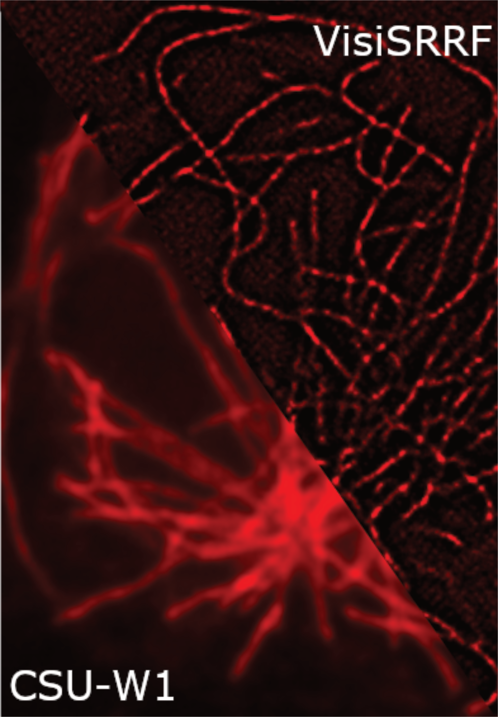
Super Resolution Radial Fluctuations (“SRRF”) is an algorithm which identifies radial fluctuations in a series of images and recalculates the origin of fluorophores by the degree of local radiality and its temporal correlation, as described by Gustaffson, N., Culley, S., Ashdown, G et al. in 2016 and Culley, S, Tosheva, K., Pereira, P., and Henriques, R. in 2018). The resolution which can be achieved with SRRF is around 70nm. The idea was developed in the lab of Ricardo Henriques and implemented in a GPU-enabled ImageJ plugin called “NanoJ-SRRF”).
ViRTEx Real‐time Experiment Control
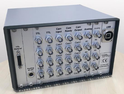
Today's technology enables new protocols for experiments employing confocal imaging, FRAP experiments, and TIRF imaging. For such protocols, which require accurate timing, Visitron Systems‘ ViRTEx technology coordinates the operation of scientific CMOS cameras, high-speed light sources such as lasers and LEDs, and piezoelectric focus motors. It communicates with cameras via TTL handshaking signals, provides TTL outputs for control of light sources and other communications, and it provides analog outputs for control of light source intensity, positioning of piezoelectric motors, and other uses.
The ViRTEx controls as many as four cameras, setting precise timing of exposure and reading the precise end of exposure. These signals provide for simultaneous acquisition from identical cameras. VisiView programs the cameras to provide the System with highest-speed acquisition.
The ViRTEx provides 16 TTL outputs for control of light source on-off, for operation of shutters, and other communications. The precise timing of of TTL signaling provides control of lighting for experiments; the high-speed reponse of lasers, LED's and galvo-based light sources allows illumination to provide for acquisition at the highest frame rates.
The ViRTEx options provide four or eight analog voltage outputs. These provide convenient control of light intensity and enable highest-speed positioning of focus or X-Y position.
Real‐time 3–D Deconvolution
The Microvolution® software delivers nearly instantaneous deconvolution by combining intelligent software programming with the power of graphics processor hardware. Confocal microscopy is an oft-used technique in biology. Deconvolution of 3–D images reduces blurring from out‐of‐focus light and enables quantitative analyses, but existing software for deconvolution is slow and expensive. The Microvolutoin option is a parallelized software module that runs about 200 times faster than other software by running on a low‐cost graphics processor board (GPU).
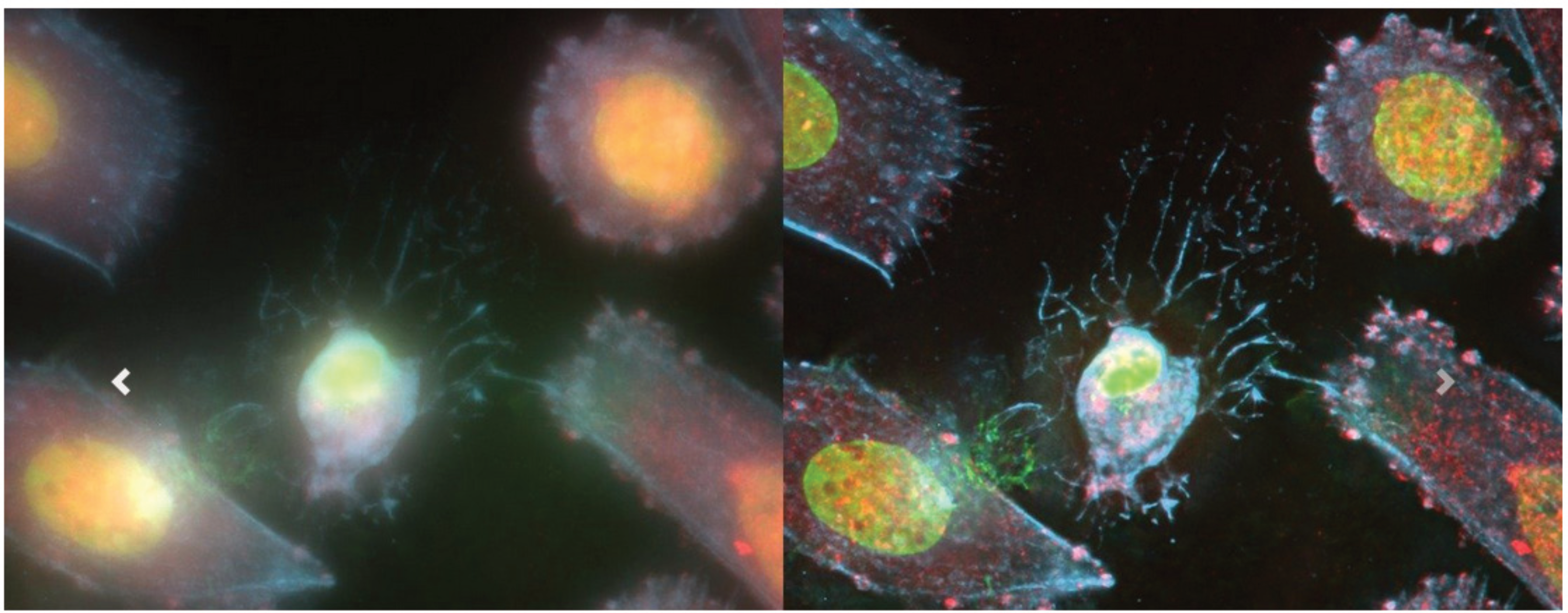
Real‐time 2–D Deconvolution
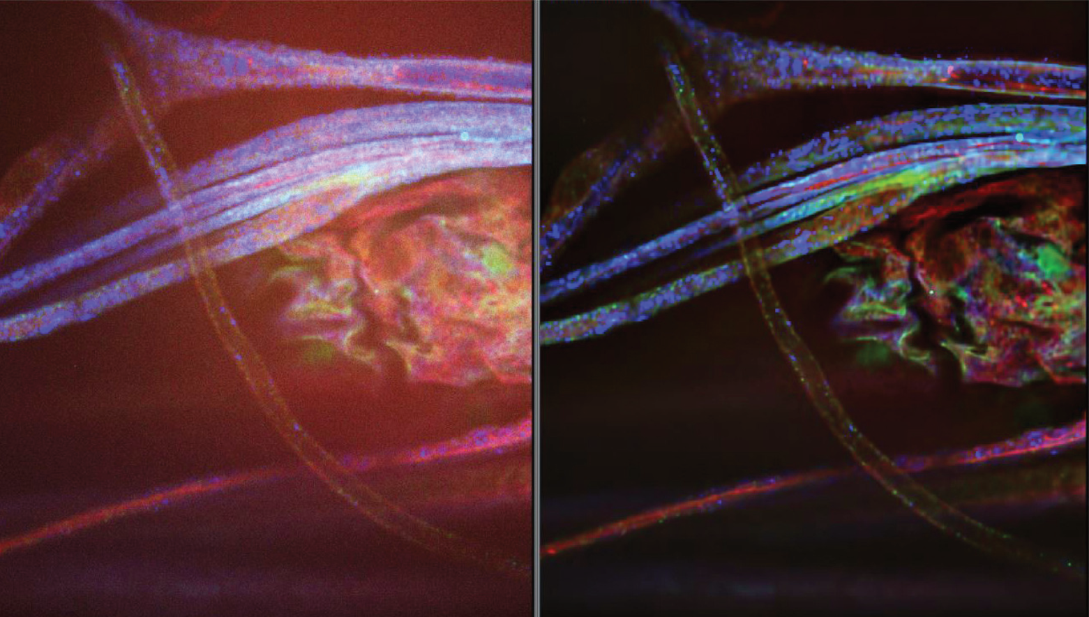
Popular widefield microscopy is a powerful optical method to visualize cellular and molecular processes involved in cells. Although fluorescent blurring from out‐of‐focus light is an limiting factor, but image deconvolution algorithms can reverse this out‐of‐focus light artifact. The 2–D real‐time deconvolution technology helps to improve resolution and contrast of a cellular image by mathematical deblurring of the image. As a result, images and fine detail become sharper while maintaining quantitative information.
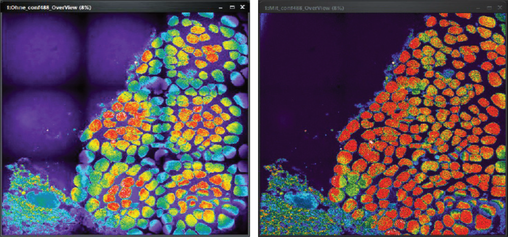
VisiView Automated Shading and Background Correction
Shading ruins the quliaty of a microscope’s images. As a consequence the intensity of objects cannot be compared reliably across a single or multiple images. Images with ugly holes in the signal are not suitable for publication. There are two main causes of shading in microscopy, number one is uneven illumination, number two is dirt.
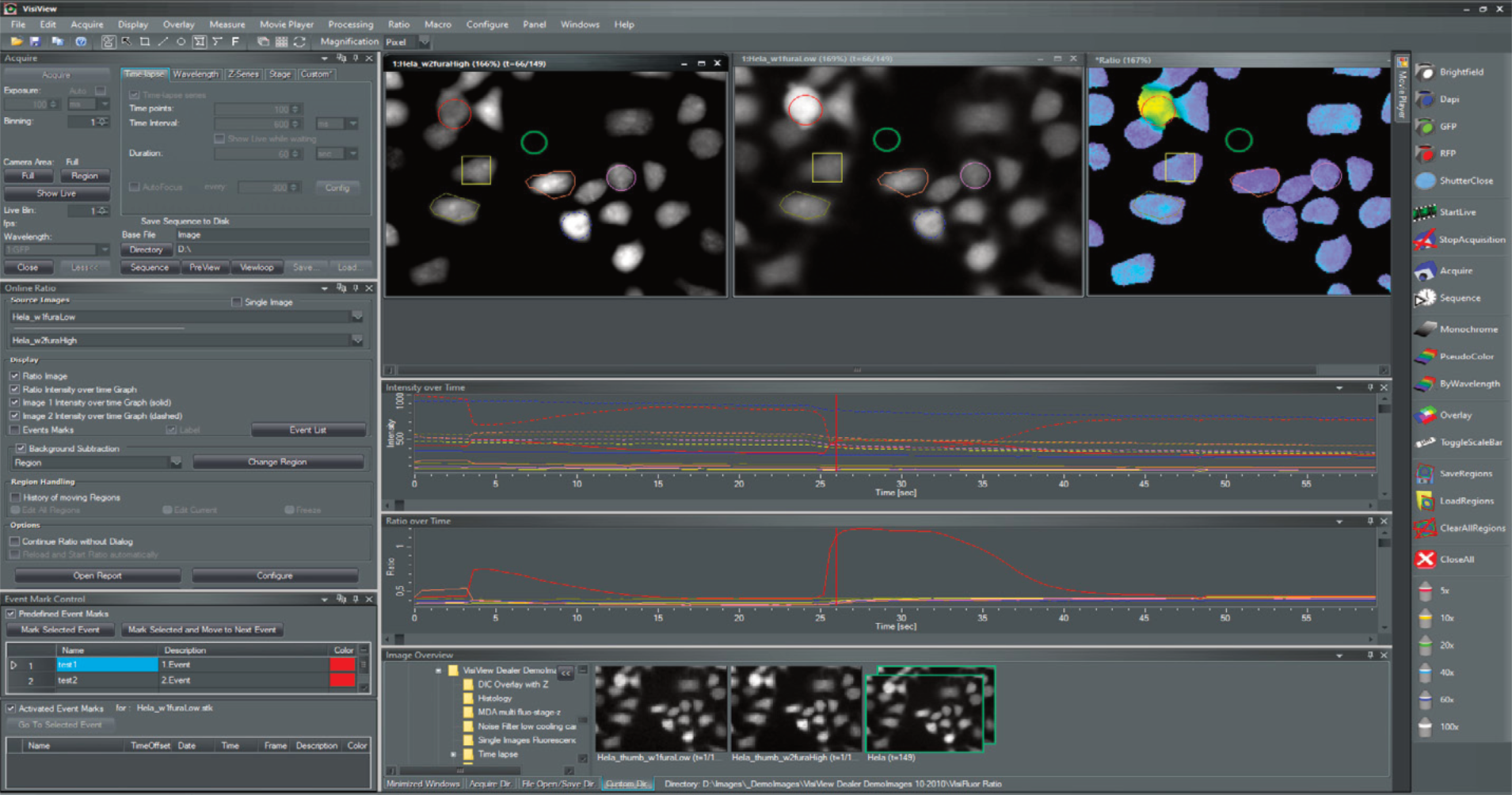
On-Line Fluorescence Ratioing Option
This option provides for high‐performance calcium, ion and FRET Imaging. Single‐wavelength images of dyes such as Calcium Green and Fluo-3 is possible for on-line image recording.
Supported Hardware
VisiView controls of a wide range of microscopy equipment:
- Color and monochrome cameras: CCD, EMCCD, scientific CMOS;
- Automated microscopes from Carl Zeiss, Leica Microsystems, Nikon Instruments, Olympus;
- Focus control, X‐Y stages, filter wheels, shutters from Ludl Electronic Products, Applied Scientfific Instrumentation, Sutter Instrument Company, and Prior Scientific;
- Piezoelectric focus control from Ludl Electronic Products, Applied Scientific Instrumentation, and other leading manufacturers;
- Standard lamps, LED light sources, and lasers;
- The VT-iSIM super-resolution confocal imager from Visitech International;
- Confocal imagers from Yokogawa Electric, Crest Optics;
- TIRF and FRAP systems: the iLas2 from Gataca Systems, the VisiTIRF and Orbital from Visitron Systems;
- Light sheet systems;
Applications
TIRF
The capture of Total Internal Reflection Fluorescence is ideal for observation of cells close to the surface of a cover slip. VisiView® supports solutions for point TIRF and ring TIRF:
- Gataca Systems iLas2 Ring TIRF and FRAP and Ablation System.
- Orbital-100 Ring TIRF System;
- Orbital-500 Ring TIRF and FRAP System;
- VisiTIRF point TIRF System;
VisiSRRF Super Resolution Algorithm
Super Resolution Radial Fluctuations (“SRRF”) is an algorithm which identifies radial fluctuations in a series of images and recalculates the origin of fluorophores by the degree of local radiality and its temporal correlation, as described by Gustaffson, N., Culley, S., Ashdown, G et al. in 2016 and Culley, S, Tosheva, K., Pereira, P., and Henriques, R. in 2018). The resolution which can be achieved with SRRF is around 70nm. The idea was developed in the lab of Ricardo Henriques and implemented in a GPU-enabled ImageJ plugin called “NanoJ-SRRF”).
Real‐time 3–D Deconvolution
The Microvolution® software delivers nearly instantaneous deconvolution by combining intelligent software programming with the power of graphics processor hardware. Confocal microscopy is an oft-used technique in biology. Deconvolution of 3–D images reduces blurring from out‐of‐focus light and enables quantitative analyses, but existing software for deconvolution is slow and expensive. The Microvolutoin option is a parallelized software module that runs about 200 times faster than other software by running on a low‐cost graphics processor board (GPU).
Real‐time 2–D Deconvolution
Popular widefield microscopy is a powerful optical method to visualize cellular and molecular processes involved in cells. Although fluorescent blurring from out‐of‐focus light is an limiting factor, but image deconvolution algorithms can reverse this out‐of‐focus light artifact. The 2–D real‐time deconvolution technology helps to improve resolution and contrast of a cellular image by mathematical deblurring of the image. As a result, images and fine detail become sharper while maintaining quantitative information.
VisiView® Automated Shading and Background Correction
Shading ruins the quliaty of a microscope’s images. As a consequence the intensity of objects cannot be compared reliably across a single or multiple images. Images with ugly holes in the signal are not suitable for publication. There are two main causes of shading in microscopy, number one is uneven illumination, number two is dirt.